
The ability for articular cartilage injuries in adults to self-repair has poor results. With new research studies and advanced technology there are a wide variety of cartilage resurfacing techniques that have the ability to improve the repair of damaged cartilage and reduce disabilities.
Of these techniques, the most advanced is articular chondrocyte implantation (ACI), which provides normal cartilage and leads to normally functioning articular cartilage.
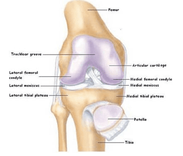
There are two main types of cartilage that are formed in the knee joint: fibrocartilage and hyaline cartilage.
Articular cartilage is composed of the hyaline cartilage, which covers the bone’s joint surfaces. The main role of articular cartilage is to ease the motion between two articular surfaces, and also provide a load-bearing surface that distributes weight and acts as a shock absorber.
Articular Cartilage Injury
As the number of adults that suffer from joint injuries continues to grow each year, treating articular cartilage damage in the knee has been a main focus over the past few years. Damage to articular cartilage could be the result of severe trauma, ligament instability, overuse, leg mal-alignment, osteochondritis, or a meniscectomy.

Cartilage Defect
Defects could develop as the result of a fall or direct impact to the knee, but most often damage occurs as the result of twisting while under a full load. The symptoms can immediately be experienced, but often times, they may not occur for several months following the initial injury. If the damaged articular surface goes untreated, it can lead to increasing cartilage degeneration, as well as osteoarthritis.
Immediately following an injury, many patients will frequently complain of acute stabbing pains and swelling that may occur 12 to 24 hours after the injury. During the first few days, bearing weight may be difficult and painful, and some patients experience the knee locking up or “giving way”.
If swelling does immediately develop, damage to internal ligaments like the anterior cruciate ligament (ACL) is likely to have occurred and needs to be examined. While there are no nonoperative specific tests to diagnose cartilage injuries, a thorough examination is valuable. The use of magnetic resonance imaging (MRI) is the most widely used tool for diagnosing cartilage injuries because it provides a visualization of surface cartilage and soft tissue structures.
Damaged articular cartilage has a limited capacity for healing naturally, which has presented major challenges for treating active individuals who wish to regain their normal functions of the knee.
ACI Repair Procedure
Over the past 50 years, many repair techniques have emerged such as the microfracture procedure, which is commonly used. Though it is an effective treatment technique with many benefits, clinical studies have shown that an ACI is a better treatment option for restoring normal joint functions.
This is because the main goal of ACI is to enable the regeneration of hyaline cartilage. Since this technique was developed, it has experienced considerable changes and is one of the most

ACI Procedure
widely used forms of treatment for defects in the knee.
The ACI technique requires two separate surgical procedures. The first procedure is performed as an outpatient surgery and includes a minimally invasive arthroscopic evaluation of the knee. The damaged area is measured and articular cartilage is removed from a surface area that is non-weight bearing, so cartilage can be cultured in the laboratory. The cartilage biopsy will then be cryo-preserved until the time of surgery.
The cartilage tissue will be thawed and cultured for the appropriate number of cell vials that are necessary for the surgery. With the second surgical procedure, the area that has been damaged is cleared of remaining cartilage and the cultured chondrocytes are then implanted.
Traditionally, this is done by injecting the cells under a periosteal flap or collagen membrane, which is then sutured onto the damaged area. This has led to some difficulties that has resulted in a second generation technique being developed.
This technique allows the chondrocytes to be cultured in a solid collagen matrix that is glued directly to the damaged area. The cells then grow inside the joint and form hard cartilage tissue that generally takes 9 to 12 months, and they continue to mature until about 24 months.
Results and Recovery

The advantages with this technique are that it significantly simplifies the implantation procedure, and this method of cell delivery allows the scaffold to act as a barrier with invasion of the graft. The best results of this procedure have been achieved with damage that is confined and localized in one specific area that is surrounded by healthy surface cartilage.
Following the procedure it is crucial that the patient follows a rehabilitation program as outlined by their Phoenix orthopedic doctor. The length of time in rehabilitation varies depending on the location of the implant. There are a variety of reasons as to why this procedure may not be successful with every patient, but long-term studies have confirmed good or excellent results with over 90 percent of the patients, and for 9 years after the procedure.
Dr. Sumit Dewanjee with FXRX is one of the top orthopedic sports medicine doctors in Phoenix and Scottsdale. He offers ACI and additional cartilage restoration procedures. This can help get patients back to sports activities and desired activities.
Dr. Dewanjee is Board Certified, Fellowship Trained and offers incredible options for knee cartilage issues, both nonoperative and operative. Call (480) 449-3979 to find out how a top Phoenix knee surgeon can help you!


